
Advanced MRI Pathology Interpretation
The MRI Report Is Not the MRI
A focused review of the actual MRI images may identify clinically significant spinal pathology that was not fully characterized or correlated with the patient’s symptoms, examination findings, and injury mechanism.
In complex spine injury cases, the actual MRI images are evaluated in conjunction with the medical history, clinical findings, prior imaging when available, and biomechanical evidence to determine what the totality of the evidence supports regarding injury, causation, and clinical significance.
MRI Image Review • Clinical-Imaging Correlation • Causation Analysis • Demonstrative Evidence
The MRI Report Is Not the MRI
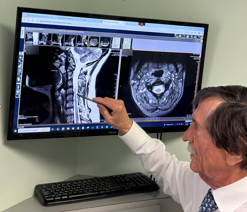
Why the Actual MRI Images Matter
A radiology report is a written interpretation and summary of an imaging study; it is not a substitute for reviewing the complete image set when clinical correlation is required.
In complex spine injury cases, the important question is not simply whether an MRI is “positive” or “negative,” but whether the imaging findings anatomically and clinically correlate with the patient’s symptoms, objective examination findings, injury mechanism, and clinical course.
The purpose of independent MRI review is not simply to identify abnormalities, but to determine whether clinically significant pathology has been fully characterized and appropriately correlated with the patient’s history, examination findings, injury mechanism, and clinical course.
Representative MRI Reviews
Representative Findings From MRI Review
The following de-identified cases illustrate how review of the actual MRI images, integrated with the clinical record, objective examination findings, and injury biomechanics, can clarify pathology relevant to diagnosis, causation, and clinical significance.
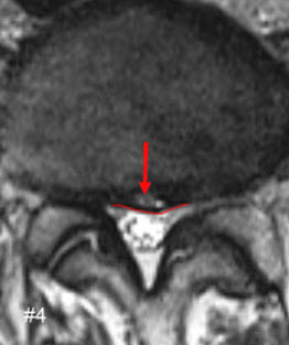
Previously Unreported Disc Herniation
The original MRI report described a disc bulge. Independent review of the actual MRI images identified a clinically significant disc herniation that had not been characterized in the original interpretation. Following re-review of the images, the interpreting radiologist authored an addendum documenting the herniation. The imaging findings were then correlated with the patient’s history, objective examination findings, clinical presentation, and documented injury mechanism, providing additional evidence relevant to diagnosis and causation.
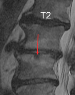
Acute Traumatic Endplate Fracture
The original MRI interpretation described reactive marrow edema associated with a Schmorl’s node and indicated that a post-traumatic etiology could not be excluded. Independent review of the actual MRI images identified features consistent with an acute traumatic disc-endplate injury. Sequential MRI demonstrated objective evolution of the endplate pathology, including Modic type 1 and subsequent Modic type 2 changes. Following review of the findings identified on each study, the interpreting radiologist authored an addendum to each MRI documenting the respective Modic changes. The sequential imaging and radiologist addenda provided objective evidence of the evolution of the traumatic disc-endplate injury and strengthened the clinical-imaging correlation regarding injury mechanism and causation.
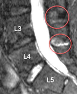
Partial Interspinous Ligament Injury
The original MRI report did not identify ligamentous injury. Independent review of the actual MRI images demonstrated increased T2 signal and edema within the L3–L4 and L4–L5 interspinous ligaments, findings consistent with partial interspinous ligament injury. The interpreting radiologist subsequently re-reviewed the study and issued an addendum confirming the ligamentous abnormality. This provided additional objective imaging support for the presence and clinical significance of the post-traumatic ligament injury.
Forensic Significance
These representative cases illustrate how independent review of the actual MRI images clarified clinically significant pathology and provided objective imaging evidence relevant to diagnosis, clinical correlation, injury mechanism, and causation.
Clinical-Imaging Correlation
The Most Important Finding Is Often the Relationship Between the Evidence

Clinical Presentation
Symptoms, neurologic findings, functional loss, and examination findings establish the clinical pattern that requires explanation.
MRI Pathology
The actual images are evaluated for disc injury, neural compromise, edema, ligament injury, endplate injury, and other structural abnormalities.
Clinical-Imaging Correlation
Imaging findings are anatomically correlated with the clinical presentation, medical history, injury mechanism, and course of care to determine their significance.
Lurie et al. compared lumbar MRI interpretations by clinical spine specialists and radiologists in patients with symptomatic disc herniation. Agreement was excellent regarding the presence and level of herniation, but radiology reports did not clearly characterize disc morphology in 42.2% of cases, and agreement regarding morphology was only fair when it was described.
These findings illustrate why review of the actual MRI images, use of standardized terminology, and clinical-imaging correlation can be important in complex spine injury cases.
A written radiology report is an important component of the diagnostic record; however, when the patient’s symptoms, examination findings, injury mechanism, or clinical course are not fully explained by the reported findings, review of the actual MRI images may provide important additional information.
The purpose of a focused MRI review is not to replace the radiologist. It is to determine whether the actual images contain clinically significant findings that require closer clinical correlation.
What the Review Examines
What the MRI Review Examines
The review is directed toward findings relevant to diagnosis, clinical correlation, injury causation, permanency, and the overall understanding of the case.
01.
Disc Pathology
Evaluation of disc protrusions, extrusions, annular disruption, endplate changes, and other findings requiring correlation with the injury mechanism and clinical presentation.
02.
Neural Pathology
Assessment of nerve root involvement, foraminal compromise, spinal canal findings, and other abnormalities that may correlate with radicular or neurologic symptoms.
03.
Trauma-Related Findings
Evaluation for edema, acute structural change, soft-tissue injury, and other imaging findings potentially relevant to the temporal relationship and mechanism of trauma.
04.
Ligament & Instability Concerns
Identification of findings that may warrant further evaluation for ligament injury or spinal instability, including correlation with dynamic imaging when clinically appropriate.
05.
Underreported or Incompletely Characterized Findings
Attention to pathology that may have been mentioned without adequate characterization, minimized as incidental, or omitted from the original report.
06.
Demonstrative Imaging
Selection and annotation of key images to help attorneys, treating providers, and jurors understand the location, character, and clinical significance of relevant pathology..
How the Review Works
01.
Review the Actual Images
The complete MRI study is reviewed rather than relying solely on the written radiology report.
02.
Compare the Radiology Report
The reported findings are compared with the pathology visible on the images.
03.
Correlate the Clinical Evidence
Symptoms, objective clinical findings, injury mechanism, relevant medical history, and clinical course are correlated with the imaging.
04.
Identify the Key Findings
The most clinically significant imaging findings are identified, characterized, and organized for clinical and forensic analysis.
05.
Explain the Significance
Relevant findings are translated into clear language and demonstrative images for attorneys, treating providers, and jurors.
When a Focused MRI Review May Be Particularly Valuable
- The radiology report does not adequately explain the patient’s symptoms
- Significant clinical findings appear inconsistent with a “mild” or “degenerative” MRI interpretation
- Causation is disputed
- Pre-existing degeneration is being used to deny traumatic injury
- Radicular symptoms are not adequately explained by the written report
- The attorney needs to understand the medical strengths and weaknesses of the imaging evidence
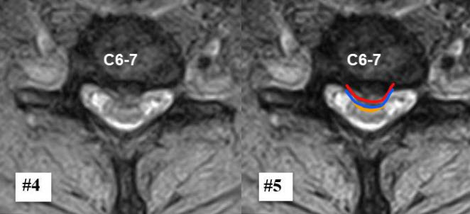
Image #4 is an axial T2 GRE image of the C6–C7 disc. Image #5 The RED LINE outlines the central disc protrusion from the annular fissure that pushes into the thecal sac defined by the BLUE LINE deforming the spinal cord as noted by the amber line.
Demonstrative Evidence
Turning Complex MRI Findings Into Clear Visual Evidence
A key MRI finding can be apparent to a trained observer yet difficult for an attorney or juror to interpret without guidance. Selected images can be enlarged, labeled, and organized to demonstrate the location, extent, and clinical significance of the pathology.
- The location of the pathology
- The involved spinal level
- The relationship to a nerve root or other structure
- Comparison with adjacent or normal anatomy
- The relationship between the imaging findings, objective examination findings, and the patient’s symptoms
Effective demonstrative evidence should clarify the medical evidence without exaggerating it.
Differentiation
From Abnormality to Medical Significance
Identifying an MRI abnormality is only the beginning. The more important forensic questions are whether the finding is clinically relevant, anatomically consistent with the patient’s presentation, temporally related to the event, and supported by the broader evidence.
Does the finding explain the patient’s symptoms and objective clinical findings?
Does it correlate with the objective examination findings?
Is it compatible with the injury mechanism?
Was the same region symptomatic or functionally impaired before the event?
Does the imaging support a structural injury or lasting functional consequence?
Can the finding be explained clearly and accurately to a jury?
CLINICAL • IMAGING • BIOMECHANICS
MRI Review Informed by Clinical Spine Experience
Peter Zilahy, DC, LAc, FPSC
- Fellow, Primary Spine Care
- Mini-Fellowship in Neuroradiology – MRI Spine
- Independent MRI Review
- Spinal Trauma & Ligament Injury
- Collision Spinal Biomechanics
More than four decades of clinical spine experience provides the context for determining whether an imaging finding is merely present—or clinically significant. MRI findings are correlated with the patient’s history, examination findings, injury mechanism, and functional loss.
Does the MRI Report Tell the Complete Story?
When the written report does not fully explain the patient’s clinical presentation, an independent review of the actual MRI images may provide important additional evidence.